
How to get rid of scars
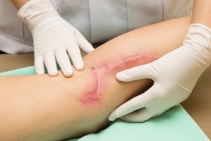
Eliminating scars through cell regeneration
is a realistic dream today for anyone undergoing skin healing. Only the knowledge of their biological evolution makes it possible to deal promptly with the aftermath of healing and deep trauma, even psychological, after damage suffered by the skin (a cut, a burn, post-surgical or post-traumatic scars).
It is the body that repairs itself by restoring an area of dermis with an equivalent tissue, albeit lacking the most peculiar characteristics of the skin (hair bulbs, sweat glands, sebaceous glands and pigmentary adnexa as well as skin lines – dermatoglyphs – that determine the uniqueness of the skin, as in fingerprints).
Regeneration or Repair which way to eliminate scars?
Cell regeneration, also known as ‘restitutio ad integrum’, occurs in fetal damage and superficial damage with complete restoration of damaged tissue with no outcome.
Tissue repair, on the other hand, takes place when the damage is deeper, leading to tissue growth that first plugs the hole present (‘loss of substance’) and then from the periphery epithelial cells called keratinocytes walk towards the centre to close the hole and make the skin waterproof.
The quality of this repair will always leave a minimal mark unlike restitutio ad integrum.
The repair of an injury varies from patient to patient because many factors come into play in the scarring process, including genetics, age, skin quality, lifestyle and exposure to sunlight.
Scars can be classified into:
- hyper- or hypopigmented scars: when they are very dark or very light compared to the surrounding skin, whose only defect is the colouring
- atrophic or depressed scars: flat but very wide and sunken scars (often occurring in patients with poorly elastic tissue or following chronic cortisone therapy)
- retracting scars: when due to their intrinsic direction they cause retraction from an aesthetic point of view if placed on the face or breasts or functional if placed on the hands or in other places where movement is essential
- hypertrophic scars when they are raised, red and painful but remain within the cut that generated them
- keloid scars are raised, painful and retracting; they are disproportionate in size to the damage that generated them; they frequently occur on the chest following heart surgery or on the earlobe as a complication of an earring hole
Evolution of the treatment and therapy of pathological scars
It is impossible to completely erase a scar but it is possible to improve its appearance. Scars are treated on patients of all ages and all over the body, while facial or acne scars are the most psychologically debilitating.
Non-invasive topical local outpatient treatments
The treatment or therapy of scars, in the first year after their onset, requires non-surgical measures such as the application of silicone-based creams, physiotherapy (by means of appropriate massages), and the application of elasto-compressive sheaths made of silicone fabric.
The objective of all these treatments is
- restore the elasticity of the collagen, which modifies the characteristics and orientation of the cells so that the scar ‘does not pull’
- compressing the scar to reduce excess microcirculation
- occlusion using silicone creams to reduce oxygen tension and suffocate them from the outside
- sun protection to prevent pathological pigmentation and reduce UV damage
- reducing scar pain and hypersensitivity
the natural goal is for a scar that is reddened in the first month to slowly regain an almost even to light-white colour and to be free of pigmentation defects.
Medical treatments to eliminate pathological scars
In the course of stabilising scars that lasts about 12 months, minimally invasive medical treatments can be set up to improve the natural evolution of scars:
In particular:
- intralesional cortisone infiltration to lower the height of a hypertrophic or chelodea scar and reduce pain and itching
- cockail infiltration of amino acid biostimulants with growth factors for atrophic scars or stretch marks that are still reddened as they evolve
- infiltration of redensifying substances underneath a depressed scar in combination with subicision to detach adhesions and give support to newly freed superficial tissue
- treatment with fractioned co2 laser to reduce dyspigmentation and smooth out scar irregularities and compact their appearance
Surgical treatment to eliminate pathological scars
Once the scar has stabilised, i.e. at least 1 year after its onset, more invasive techniques can be used for treatment:
- flat or atrophic scars: surgical revision, i.e. to change the position, shape and course, the entire scar is surgically removed with the margins included, which are sutured again in the most accurate and least reactive manner possible.
- retracting scars: surgical revision and V-Y plastic or plastic lengthening- implantation of skin expanders (especially on the scalp) Z or W plastic or transposition of flaps
- fractioned CO2 ablative laser causing soft dermabrasion and collagen regeneration (Slim II – Mixto Pro – Lasering)
- subcision with lipostructure, subcutaneous nanofat grafting with the SNIF technique ( sharp needle intradermal fat) achieves regeneration of fibrous tissue due to the gain of elasticity from newly transplanted adipose stem cells while subcision ( rigottomie) creates a release of tension giving the stem cells room to graft.
- depressed scars ( also many acne scars or deep stretch marks, also skin dimpling from gluteal cellulite ) :
- fractioned CO2 ablative laser causing soft dermabrasion and collagen regeneration (Slim II – Mixto Pro – Lasering)
- subcision with lipostructure, subcutaneous nanofat grafting with the SNIF (sharp needle intradermal fat) technique achieves regeneration of the fibrous tissue thanks to the gain of elasticity from newly transplanted adipose stem cells, while subcision (rigottomie) creates a release of tension, giving the stem cells room to graft.
- hypertrophic or keloid scars it must be said that simple removal of a keloid is absolutely forbidden because the risk of recurrence is almost certain in the absence of complementary therapy.
- therapy by infiltration of slow-release cortisone (triamcinolone acetonide-kenacort), which relieves pain, reduces swelling but does not allow cosmetic improvement but only functional improvement
- surgical revision with complementary radiotherapy by means of external beams or brachytherapy (after removal of the scar, adjuvant treatment is carried out within 72 hours); the most frequent sites are the auricular lobe and the presternal region, which are often unpredictably predisposed to the formation of keloids or hypertrophic scars, with success rates of over 60%.