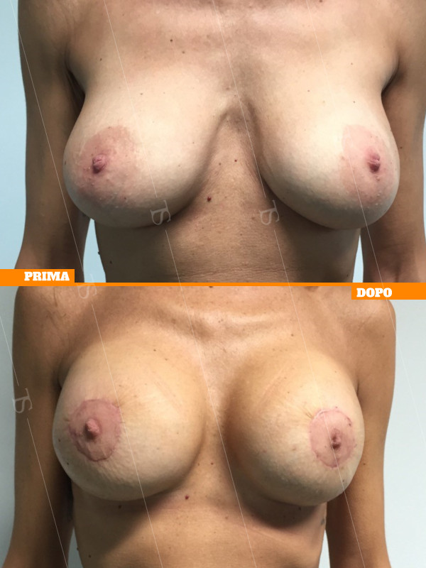

Breast implant replacement or Secondary breast augmentation
One of the most requested cosmetic surgery procedures in recent times concerns the replacement of breast implants, through a procedure called Secondary Mastoplasty.
The procedure may involve the repositioning of tissues, the correction of defects that the previous surgery did not correct or imperfections that occurred years after the first implant.
Often Dr. Tommaso Savoia is asked to replace the prosthesis in order to increase (or decrease) its volume.
This is an intervention that presents several pitfalls but that in some cases is not to be missed, especially when there is deterioration or deformation of the old prosthesis.
Breast implants have a history that dates back to March 1962 when the plastic surgeons Cronin and Gerow implanted the first pair of silicone implants.
Over the years, implants have undergone continuous evaluation and re-evaluation to ensure their safety. We have also passed to prostheses filled with saline, hydrogel, polyurethane and soybean oil to return with safety and quality to cohesive silicone that allows even in cases of rupture that there is no leakage.
The main causes and related solutions that lead a patient to undergo a secondary mastoplasty are:
- Aesthetic changes in shape and volume: prosthesis replacement, possible capsuloplasty and mastopexy.
- hardening of the breast or capsular contracture: capsulectomy and prosthesis replacement.
- prosthesis rupture: prosthesis replacement.
- breast ptosis: reduction mammaplasty and implantation of small prosthesis.
- prosthesis displacement or dislocation: capsulotomy and capsuloplasty.
- hardening from radiotherapy or breast reconstruction outcomes: lipofilling or capsulectomy and prosthesis replacement or reconstruction with muscle flaps.
The patient who suspects a rupture (sometimes does not even realize it) feels a lesser turgidity of her breast to the touch because, being accustomed to a certain consistency only she perceives the difference not so much compared to the contralateral breast but especially according to the previous days or months.
The first investigation to be carried out is the ultrasound that investigates both the glandular tissue and the wall of the prosthesis, but it is important that the operator focuses only on the breast because this examination is useful only if the operator is specialized in the field.
Prenota una visita
Click HereSecondary breast surgery and breast implant replacement
Secondary breast surgery is a procedure to revise or replace breast implants following a previous breast augmentation.
When deciding to have breast implants fitted for the first time, it is important to bear in mind that further surgery will almost certainly be required within 10–20 years.
The reasons leading to revision breast augmentation surgery are:
– painful or ‘marble-like’ capsular contracture classified as Becker grade 3 or 4
– displacement or dislocation of the breast implants
– a desire for a greater or lesser volume due to changes related to weight, weight loss, pregnancies or personal expectations.
– rupture of a previous implant, which may be non-urgent, as in the case of an intracapsular rupture, or more urgent if the implant is old and the rupture is considered extracapsular with gel bleeding (leakage of silicone material beyond the capsule into the lymph nodes)
– Reaction to the implant, such as recurrent seroma or a diagnosis of anaplastic lymphoma associated with textured implants (BIA-ALCL)
– Immediate or delayed haematoma due to excess blood, necessitating opening of the wound with a risk of infection
– Prosthesis infection due to immunosuppression, or sepsis originating from within
– Breast ptosis or prosthesis displacement due to changes in the anatomical conditions
– Prosthesis rejection, i.e. the patient has suffered various complications and wishes to have the prosthesis permanently removed without reimplantation.
Breast implant check-up prior to secondary breast augmentation
The guidelines of the American College of Radiology recommend that a new ultrasound scan and mammogram be carried out both before and after the first year following surgery. Indeed, after 12 months, the internal surgical scars are no longer visible on radiological images, and it is therefore possible to assess the condition of the mammary gland and the implants more effectively.
Patients should undergo an MRI scan every 3 or 4 years, and more frequently in patients at high risk of cancer due to family history or those with fibrocystic breast disease, which is a physiological condition that is more difficult to assess.
To dispel a myth, we must remember that 10 years is not the lifespan of the implant after which it ‘expires’, but rather the most common duration of the manufacturer’s warranty. Implants should only be replaced if there are clinical reasons, such as rupture, displacement or painful capsular contracture. Other reasons are personal ones, which often lead the patient to request a larger volume because, after 12 months, the effect—whilst still pleasing—is less pronounced than after 1 month due to physiological settling.
The actual lifespan of a breast implant varies considerably, as it depends on individual factors relating to how the patient’s tissue reacts to the implant or to random manufacturing defects. In clinical experience, implants have been found to have ruptured after 2 years
and implants that remain perfectly intact even after 28 years.
The key recommendation is to undergo regular ultrasound or mammography screening for cancer prevention and to monitor the condition of the implants.
For diagnostic confirmation or if a rupture of the implant is suspected, a magnetic resonance imaging (MRI) scan is carried out, which will allow for a detailed examination of the implant and the capsule surrounding it.
It is important to note that, over the course of a lifetime, the patient will certainly need to have the implants replaced and therefore undergo a second operation.
How the capsule surrounding the breast implant forms
After implantation, the implant stimulates the formation of a layer of fibrous connective tissue that isolates it from the rest of the body; this is known as the periprosthetic capsule. It is a physiological and natural part of the healing process that the surgeon intends to occur in order to keep the implant in the desired position established at the end of the operation, which also serves to protect it from infection.
In fact, the first month following surgery is very important for the stability of the long-term result, and the surgeon may recommend a specific type of massage or the use of an elastic band to stabilise the implant, soften it and better define the pocket of the new breast.
The tissue covering the implant consists of fibrous collagenous connective tissue, fibrous cells known as fibroblasts, and other cells known as myofibroblasts, which are responsible for creating varying degrees of tension. This capsule can change and stabilise over the course of the first year following breast augmentation.
Baker’s classification of capsular contracture
Baker’s classification describes the extent of the connective tissue reaction produced by the body in response to the implant.
Grades I and II on the scale correspond to breasts with natural and physiological results, as they are characterised by a soft scar tissue capsule that the patient does not even notice. Grade III, on the other hand, is a condition in which the patient feels the capsule is stiffer and perceives the implant as firmer and sometimes painful. In Grade IV, the capsule surrounding the implant is so thick and hard that it causes severe pain and a sensation of pressure in the chest due to compression of the sensory nerves. Furthermore, the implants are displaced upwards and constricted concentrically, making them appear smaller and more oval in shape.
This clinical condition frequently occurs in patients who have undergone surgery for breast cancer following radiotherapy or a very prolonged breast expansion procedure.
The causes of capsular contracture are:
individual and unpredictable, often with asymmetrical results
a subclinical infection, i.e. contamination that can lead to hardening of the implant due to the inflammatory process. Some risk factors include bacteraemia in patients with weakened immune systems, smokers, or those with poor dental and oral hygiene.
an untreated haematoma, i.e. blood remaining in contact with the implant, which prolongs the healing time and causes inflammation
a seroma, i.e. the formation of a yellowish fluid within the implant pocket, which may initially cause the implant to shift; even after its removal or reabsorption, it leaves behind a stiffer capsule
radiotherapy treatments for breast cancer
subglandular implantation has a significantly higher rate of developing capsular contracture than submuscular implantation; furthermore, the outline of the implant is more visible in slim or elderly patients.
periareolar and axillary incisions are associated with a higher incidence of capsular contracture than the inframammary fold incision
Medical treatments and massage
Capsular contracture can, in some non-severe cases, be treated by means of targeted massage of the capsule to soften it and stretch the collagen fibres. This treatment is suitable in situations where the patient only feels tension on palpation and discomfort, but not yet pain, and the implant is not deformed but, at most, simply feels firmer in the upper pole. In this case, surgery may even be avoided.
This is often combined with ultrasound sessions, as described in the Aspen protocol for capsular breast contracture.
There have been studies – though not yet certified – on the efficacy of anti-inflammatory drugs based on leukotriene inhibitors, such as those used to prevent asthma relapses (Montelukast – Singulair), at a dose of 10 mg per day. This protocol has been the subject of some scientific publications that have raised hopes, but it is not yet an established treatment.
Surgical Solutions for Capsular Contracture Following Secondary Breast Augmentation
In cases where pain is present alongside displacement, surgery will be necessary, involving the removal of the implants and the complete excision of the surrounding capsule (radical capsulectomy – en bloc capsulectomy).
Alternatively, in less severe cases, the surgeon may make precise incisions in an orderly fashion. If we imagine the breast as a half-sphere, incisions are made along the equator and along the meridians to expand the space housing the implant (radial and circumferential capsulotomy).
We refer to capsuloplasty when we use capsular flaps to alter the shape of the pocket containing the implant, or when we need to thicken the lining, or when we need to narrow a pocket that is too wide in cases where the implant has shifted.
Surgical solutions regarding implants and the implant pocket
Replacing the implant with a new one is essential whenever the implant pocket is compromised.
In the case of capsular contracture, where possible, it is advisable to change the implant placement from subglandular to submuscular, for example, or vice versa.
In some cases, it is recommended to replace silicone implants with implants containing silicone but coated in polyurethane, as a significantly lower incidence of capsular contracture has been observed. In fact, the polyurethane coating on the implants adheres more firmly and is thought to reduce stimulation of the fibrous tissue of the capsule, whilst it is gradually reabsorbed and integrated into the surrounding tissues over a period of 10 years.
In cases of contracture that are not severe in terms of shape but are particularly painful, or where there are superficial irregularities (rippling), regenerative treatment using adipose tissue grafting (lipofilling) is recommended to reduce superficial inflammation. The aim is to help soften the capsule, reduce pain caused by nerve compression and smooth out superficial irregularities. This treatment is minimally invasive but often requires two or three sessions, each spaced three or four months apart.
In more severe cases within the field of post-cancer breast reconstructive surgery, where the implant is not tolerated and forms a very painful capsule, muscle flaps are used to cover the implant (latissimus dorsi muscle flap) or even replace it (rectus abdominis muscle flap).
The surgeon therefore finds themselves operating on a patient who has already undergone surgery; for this reason, it is important to consult an experienced surgeon. Before proceeding with secondary breast augmentation, Dr Savoia always carries out a careful assessment of each individual case to understand the patient’s problems and expectations and ultimately deliver the desired result.